During Mr. A’s most recent ultrasound exam, doctors noticed a slight increase in the size of a previously known complex cyst in his right kidney. A simple cyst would normally appear well-rounded and have clearly defined boarders and fluid with a consistency similar to that of water. By comparison, a complex cyst may not be as well rounded, and often has ill-defined borders and contents other than water-like fluid. Because complex cysts more often turn out to be malignant, they are more closely monitored. And because of Mr. A’s known cirrhosis, doctors were concerned that the cyst may indicate the presence of either a primary kidney cancer or metastatic disease from an undiagnosed HCC.
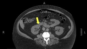
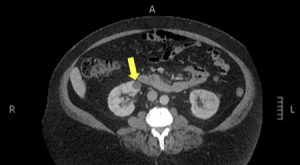
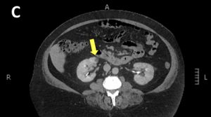
A CT scan was recommended to further evaluate the complex cyst and assess whether the mass is enhancing (i.e., receiving a portion of the body’s blood flow), which can be worrisome. However, the CT images were inconclusive due to the patient’s body habitus and the specific characteristics of the cyst.
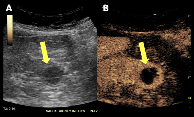
The radiologists then turned to contrast enhanced ultrasound (CEUS) to further evaluate the cyst. In particular, doctors wanted to use CEUS to determine whether the cyst had grown and whether blood was flowing to it.
The CEUS scan was performed after 6 months, and the procedure took approximately 15 minutes. The radiologists used an FDA-approved ultrasound contrast agent with microbubbles to evaluate the suspicious cyst in real-time without ionizing radiation or iodine, which may be associated with risk of damage to the kidneys.
CEUS revealed that the cyst did not have any enhancement and was indeed a simple cyst.
Because of CEUS, this cyst no longer requires follow up. Mr. A will continue to visit his doctor and have annual ultrasound surveillance of his liver.