
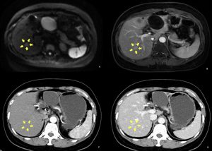
Doctors there performed contrast-enhanced liver CT and MRI. The MRI images showed an approximately 1.5cm-sized lesion with restricted diffusion and sustained enhancement in segment 6 of the liver (Fig. 1A, 1B). The CT scan showed a suspicious nodule with inhomogeneous mild enhancement (Fig. 1C, 1D). Given these findings, the patient’s liver mass was considered as liver metastasis. Then she was hospitalized for further treatment.
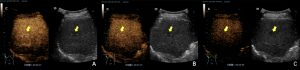
To further characterize the liver mass, a contrast-enhanced ultrasound (CEUS) examination was obtained. CEUS demonstrated iso-enhancement of the lesion on all contrast phases (Fig. 2) compared to surrounding parenchyma. This kind of contrast enhancement pattern was typical for a benign portion of heterogeneous fatty liver.
In the end, a liver biopsy was carried out to confirm. The histologic evaluation revealed modest fatty infiltration along with localized hepatocellular hyperplasia. No tumor cells were discovered when combined with the immunohistochemical staining data. This ruled out the more concerning diagnosis of metastasis and supported the diagnosis of heterogeneous fatty liver.
Thanks to the CEUS exam, Mrs. H will be able to avoid unnecessary operations and receive better care. She is still being followed with abdominal US and chest CT. However, there is no need to concern about liver metastasis given the CEUS results and the biopsy’s demonstrated absence of tumor cells.