
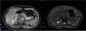
At the time of his initial checkup, Mr. Hua was not experiencing abdominal bloating, nausea, or other abdominal pain or discomfort, and he had no significant medical history or family medical history. Following the ultrasound, physicians performed a gadolinium-enhanced liver magnetic resonance imaging (MRI) and found a 3.5 x 3.0cm liver mass (Figure 1).
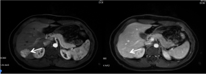
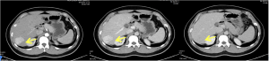
Given these findings, the patient’s liver mass was considered as hepatic cancerous (HCC) and the patient was referred to our hospital for surgical treatment. All of his laboratory data and tumor markers were at normal level. A follow up contrast enhanced liver CT (Figure 2) scan was performed to future confirm the result. The CT scan showed a hypo-intense mass in the segment 6 of the liver, which could have been considered HCC.
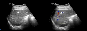
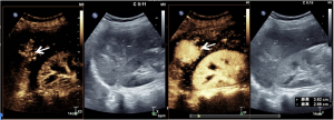
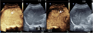
To further characterize the liver mass, conventional ultrasound and contrast enhanced ultrasound (CEUS) scans were administered. Initial gray scale images showed a 3.9 x 4.3cm hyperechoic heterogeneity mass in the segment VI of the liver, with feeding blood flow on Doppler ultrasound (Figure 3). After administration of the ultrasound contrast agent, the mass showed centrifugal enhancement in early arterial phase and sustain hyperenhancement compared to normal liver parenchyma during three phases (Figure 4).
Given these findings and the pattern of contrast enhancement, the liver lesion was considered as a common noncancerous condition called focal nodular hyperplasia (FNH).
However, since both CT and MRI considered the liver mass as HCC, the patient underwent laparoscopic surgery. Fortunately, the histological examination showed that the liver mass was FNH, as indicated by CEUS, and Mr. Hua has done well since surgery.
As Mr. Hua’s experience shows, CEUS can depict real-time blood flow and provide more valuable diagnostic information than MRI and CT.