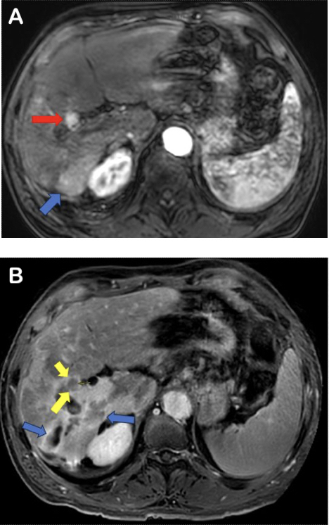
His initial diagnosis was made two years ago, after which he underwent a series of targeted treatments including chemoembolization and selective internal radiation therapy. Recently, on a routine follow-up MRI scan, a new liver lesion was identified with features suggestive but not conclusive of recurrent cancer. In some cases, previous treatment areas can exhibit confounding imaging characteristics, which complicate the evaluation for recurring malignancy.
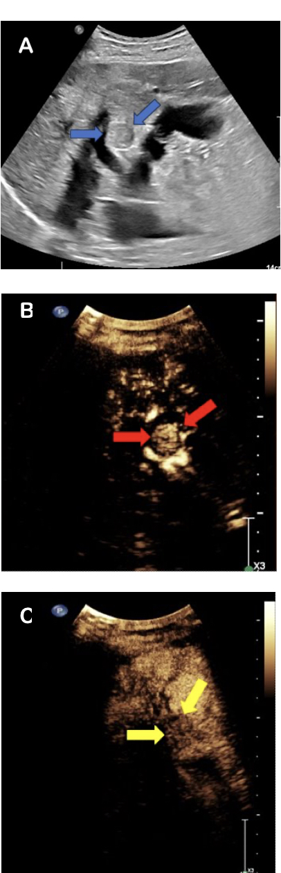
Given these findings, physicians recommended a contrast-enhanced ultrasound (CEUS) scan to assess feasibility of radiofrequency ablation, a minimally invasive treatment technique. CEUS offers a quick and precise problem-solving tool, capable of diagnosing primary HCC’s, confirming suspicious nodules as in this case, or finding masses that were not seen confidently on ultrasound without contrast alone.
In under 10 minutes, the radiologists were able to use microbubble ultrasound contrast agents and conventional ultrasound equipment to evaluate the suspicious nodule in real-time without the risk of ionizing radiation or toxicity to the kidneys – which was especially important because of Mr. H’s chronic kidney disease.
CEUS revealed the small target which measured under 2 cm and was able to distinguish it from nearby treatment areas, confirming the diagnosis of recurrent cancer. Additionally, CEUS effectively localized the new tumor in relation to important nearby structures, including veins and bile ducts. Ultimately, CEUS provided a precise diagnosis with no harm to Mr. H, while also influencing the treatment options that were available. This gave physicians confidence in identifying the nodule as the correct target in this technically challenging liver area, given the surrounding changes from prior treatments.
Thanks to CEUS, Mr. H had the benefit of a precise, cost-effective diagnostic imaging tool, distinguished by its high level of safety and its availability for patients with kidney disease. Thanks to the invaluable insights provided by CEUS, Mr. H is now scheduled for another chemoembolization session, after which CEUS can continue to be used to assess his response to local-regional therapy at minimal relative financial cost.