Labs: the inflammatory markers do not detect inflammation.
39-y/o male patient came to our emergency department due to sudden pain in the lower part of the abdomen.
A baseline ultrasound examination was recommended for follow-up.
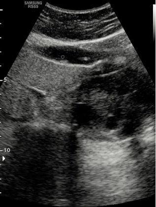
Echographic findings: In the right liver lobe, conventional sonography delineates a 6 cm hypoechoic lesion with cystic areas.
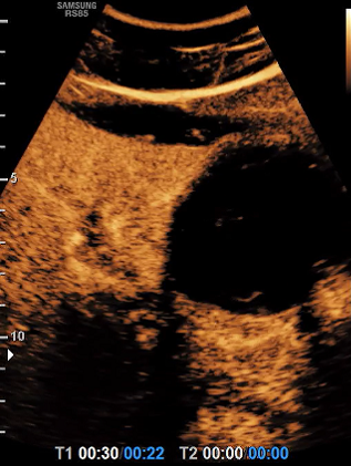
After the native ultrasound examination, CEUS was recommended. The lesion does not show any contrast uptake.
Grey-Scale examination
Color doppler examination
Choose your diagnosis:
CEUS arterial examination
CEUS portal venous phase examination
CEUS late phase examination
Choose the final diagnosis:
#echinococcosis alveolaris.
The serology test confirmed the diagnosis.
CEUS depicts a safe method for the evaluation of echinococcal liver disease. In addition to serological tests and grey scale ultrasound, CEUS imaging could be integrated as an easily accessible tool helping to describe hypervascularization as a sonomorphological correlate for active perilesional inflammation of echinococcal manifestations.
The information, references and opinions provided on this website do not constitute the offering of medical advice or create a physician-patient relationship between any one or more of (a) the International Contrast Ultrasound Society (ICUS), its directors and members, and the contributors to this website, and (b) you, your patients, and any other party. ICUS makes no express or implied warranties regarding the accuracy or completeness of the information, references and opinions on this website, and ICUS shall not be liable to you, your patients, or anyone else with respect to any medical decision made, or any action taken or not taken, in direct or indirect reliance on this information.


International Contrast Ultrasound Society © 2025. All Rights Reserved.