The CT scan showed a spontaneous, contained bleed around his right kidney with no obvious source (Figure 1). These types of bleeds can be caused by both benign and malignant renal tumors, vascular abnormalities, and inflammatory disorders. To further investigate, an abdominal MRI was obtained. The MRI images showed a suspicious nodule adjacent to the bleed that was not completely characterized (Figure 2). This finding was concerning for a mass, however it remained indeterminate.
The radiologists suggested to Mr. S and his urologist to consider pursuing a contrast enhanced ultrasound (CEUS) to better characterize the mass and provide some clarity to the situation. With the use of safe gas-filled microbubbles, contrast enhanced ultrasound can provide great visualization of organs and blood vessels. It’s a safe and quick imaging technique that adds accurate diagnostic information at a low cost.
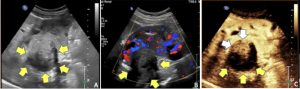
A joint decision was made to undergo the contrast enhanced ultrasound. Within seconds after injection of contrast, microbubbles illuminated the kidney and its vasculature. The mass was confirmed and strong contrast enhancement was seen (Figure 3). This finding was highly concerning for renal cell carcinoma, a malignant tumor of the kidney. The tumor had bled earlier but no longer showed any active bleeding during the imaging exams. By providing fast and essential diagnostic information about the mass, the patient was able to expedite further care and treatment.
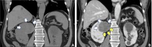
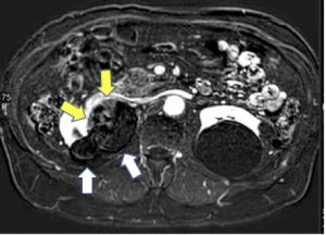
Figure 2. MRI reveals a bilobed hematoma within the right kidney (white arrows). The subtle enhancement seen on CT is better shown as a bright nodular enhancing component on the early contrast phase (yellow arrows).