Resident, Department of Radiology, University of Washington, Seattle, WA
Share
CEUS Excludes Kidney Mass
Ms. S is a 64-year old Cambodian woman with chronic active hepatitis B currently being treated with tenofivir alafenamide fumarate, a medication to treat the viral infection of her liver. She has no radiologic or laboratory evidence of cirrhosis and has been undergoing screening abdominal ultrasounds every six months according to international guidelines due to her increased risk for liver cancer given the diagnosis of hepatitis B, which have been unremarkable.
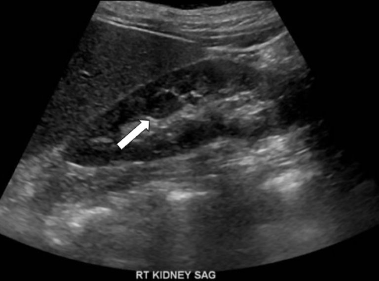
Her most recent right upper quadrant ultrasound demonstrated as an incidental finding a newly appeared solid oval area in the midportion of the right kidney without internal vascularity. This finding was indeterminate on the current study and thought to represent a complex cyst, solid mass, or pseudo-mass, which is not a real pathologic finding, but just an unusual appearance of normal tissue. Further evaluation with contrast-enhanced ultrasound was recommended.
Figure 1: Ultrasound of the right kidney in sagittal orientation (along its long axis) demonstrates a well-circumscribed nodular solid appearing lesion in the interpolar (mid) region of the right kidney, isoechoic to the adjacent renal cortex (arrow).
Figure 2: Color Doppler ultrasound of the same area of the right kidney shows there is no increased vascular flow except for normal blood vessels (red dot close to arrow).
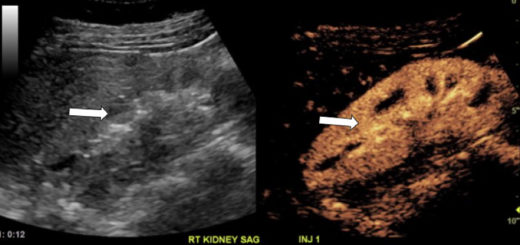
Two weeks later, a focused contrast-enhanced ultrasound of the right kidney was performed. Before the administration of IV contrast, the lesion was again seen in the interpolar region of the kidney, unchanged from the prior exam. Real-time sonographic images of the right kidney during and immediately after contrast administration showed that this area enhanced identically to the adjacent (renal cortex) kidney tissue during the entire exam. There was no evidence of washout or architectural distortion. Therefore, the previously indeterminate right renal lesion was a prominent normal area of the renal parenchyma rather than a complex cyst or solid mass mimicking a tumor on conventional ultrasound (so-called pseudo-mass). This information was immediately conveyed to the patient.
Figure 3: Sagittal contrast-enhanced ultrasound images of the right kidney with conventional mode ultrasound on the left and ultrasound optimized to show only the microbubble contrast agent of the same area simultaneously on the right: exam redemonstrates the isoechoic area in the interpolar region of the right kidney (arrow on left image), which is unchanged from the prior exam. This area enhances homogenously and identically to the rest of the kidney during the five minutes of observation after contrast injection: image on the right taken 12 seconds after contrast administration shows that the nodule is not discernible from the surrounding kidney tissue (arrow), which is typical for a normal portion of kidney.
In this scenario, CEUS provided immediate peace of mind to Ms. S and prevented further unnecessary workup of the presumed renal abnormality with multiphase CT/MRI or biopsy. The definitive diagnosis was able to be made with a noninvasive study and the immediate reassuring diagnosis was conveyed to the patient, who was relieved that she did not have a kidney tumor.