Resident, Department of Radiology, University of Washington, Seattle, WA
Share
CEUS allows patient to avoid expensive MRI and risky biopsy
Mr. Y is a 56-year-old father of two. During a recent routine visit to his doctor, he said he felt good and appeared to be healthy. Unfortunately, a digital rectal exam proved to be abnormal and Mr. Y’s prostate specific antigen blood count was elevated. A subsequent biopsy showed signs of prostate cancer.
To evaluate the size of Mr. Y’s prostate cancer and how far it may have spread, he underwent a bone scan and a pelvic MRI. Fortunately, the bone scan demonstrated no metastatic lesions in his skeleton. However, the pelvic MRI showed that two lymph nodes were slightly enlarged.
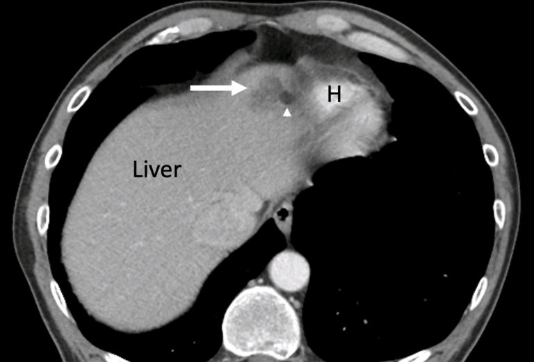
Mr. Y then underwent a CT of his abdomen and pelvis so physicians could determine whether other metastatic disease was present. The abdominal CT showed a 2.5cm mass-like, slightly ill-defined structure in the liver, which was not completely characterized.
Given the diagnosis of cancer in the prostate, it was unclear whether this structure represented prostate cancer that had spread to the liver or something else. And, since the structure was located relatively close to the heart, physicians were concerned that a biopsy would be risky and difficult to perform. Instead, they recommended a second MRI scan, this one focused on the liver and performed with and without contrast.
Mr. Y. was, however, reluctant to undergo another expensive MRI scan or a biopsy. After consultation with his urologists, he was referred for a contrast-enhanced ultrasound (CEUS) scan of the liver.
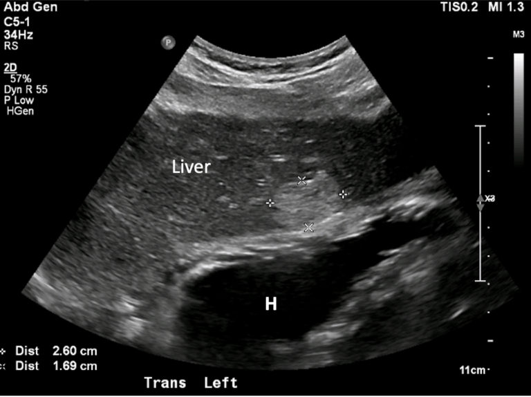
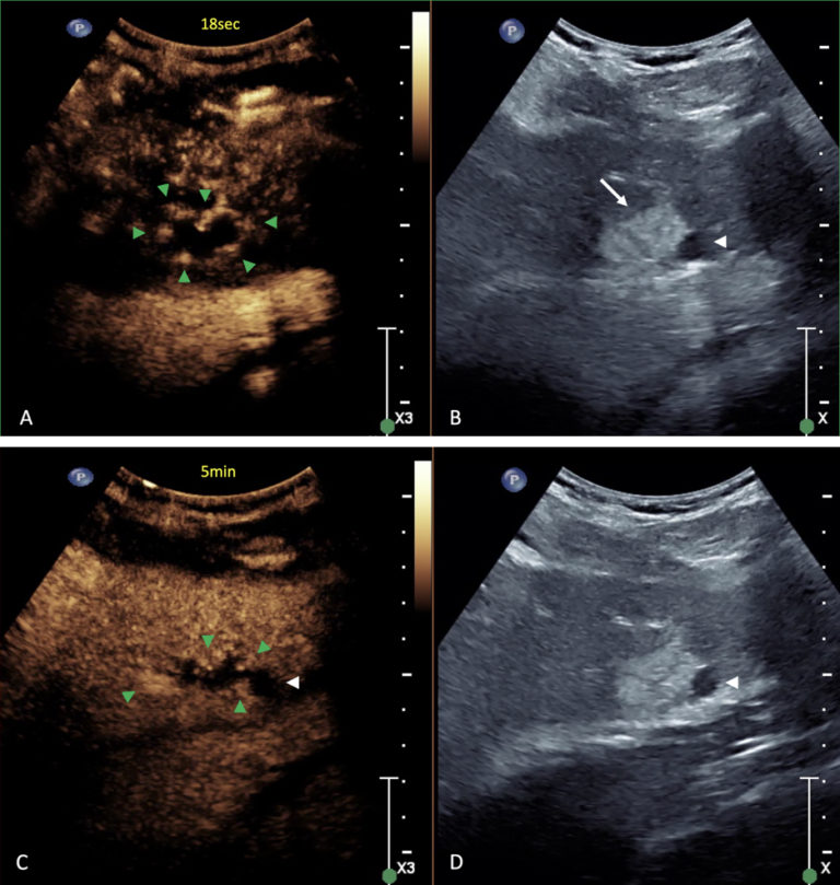
The CEUS scan was relatively inexpensive and performed without delay. It showed an echogenic mass in the left lobe of the liver corresponding to the finding on CT with peripheral nodular contrast enhancement and gradual fill-in of contrast over the observation period of five minutes towards the center. Centrally however no microbubbles were seen.
Fortunately for Mr. Y, these CEUS images were consistent with a hemangioma with central clots (thrombus) — a very common benign abnormality of blood vessels. The central clots had been difficult to diagnose using a limited CT, which had been performed with only one contrast phase for screening purposes.
Mr. Y learned the good news at the end of his CEUS exam, and was relieved to hear that the nodule in his liver was benign and not a metastasis.
Thanks to a relatively quick and easy CEUS exam, Mr. Y received a reliable diagnosis and could begin treatment of his prostate cancer without further delay.
CT of the abdomen shows an irregular focal hypodense abnormality (arrow) in the left lobe of the liver, close to the heart (H). A small cyst (arrowhead) is located next to the lesion
Transverse ultrasound of the left lobe of the liver shows a 2.6 x 1.7cm echogenic mass lesion (outlined by distance markers) in the liver corresponding to the atypical finding on CT. Note close proximity of the lesion to the heart (H). The small cyst is not shown on this image.
Contrast-enhanced ultrasound of the mass (arrow) in the left liver lobe, with contrast mode optimized to show only the microbubble contrast agent on the left (A, C), and conventional mode on the right (B, D): the lesion shows nodular peripheral enhancement 18sec after intravenous injection of the agent (green arrowheads in A) which slowly but incompletely fills in after 5minutes (green arrowheads in C) leaving a black central void (A and C), typical for a benign centrally thrombosed hemangioma. Note that the small simple cyst (white arrowhead) does not enhance with contrast, which is expected for a cyst.