curated by Dr Dirk Clevert
February 7, 2022

The B-mode examination demarcates a heterogeneous lesion in the right lobe of the liver, measuring 10.2 x 9.0 cm

The color Doppler examination shows no internal blood flow signal.

Labs: The inflammatory markers do not detect inflammation

39-y/o male patient with abdominal pain underwent abdominal CT, where a liver lesion was detected.

Findings:

After intravenous contrast agent administration, the lesion shows no contrast uptake during the entire examination.

The serological test revealed the diagnosis of Echinococcus (Hydatid disease).

What is your diagnosis?

Unenhanced Images

Choose your diagnosis:

Did your diagnosis change?
Enhanced Images

Choose the final diagnosis:

Results

Case5_before Cae5_after

Diagnosis

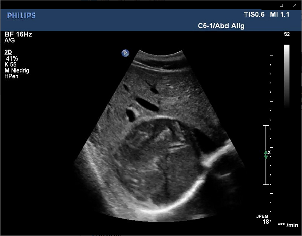
Depending on the echinococcal manifestations, patients can later develop unspecific symptoms as fatigue, abdominal pain and may present with elevated transaminases, jaundice and hepatomegaly.

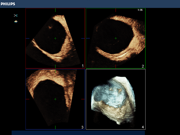
CEUS depicts a safe method for the evaluation of echinococcal liver disease. In addition to serological tests and grey scale ultrasound, CEUS imaging could be integrated as an easily accessible tool helping to describe hypervascularization as a sonomorphological correlate for active perilesional inflammation of echinococcal manifestations.

Previous
Next

Be Part of our Community

ICUS Membership is free.

ICUS Connect

FREE
VIEW