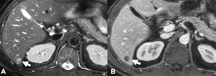
A follow up CT scan was performed to screen for other masses. It showed an indeterminate small 1 cm mass in the right lower aspect of the liver. Then a follow up MR exam was performed for further evaluation and to exclude more smaller masses that may not be well visible with CT. The MR scan did not show more masses but showed targetoid appearance of the known liver mass without showing all the imaging features of an epithelioid hemangioendothelioma. With these inconclusive findings, an ultrasound guided biopsy was needed.
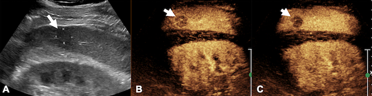
To obtain more convincing preceding evidence, physicians turned to contrast enhanced ultrasound (CEUS), which could be performed as a simple bedside procedure on the same day as the biopsy. The CEUS scan showed the tumor much better than ultrasound alone, making the biopsy safe and easier to perform. In addition, “early washout” of the contrast agent was observed, indicating that the hepatic mass was indeed malignant.
This CEUS scan conclusively justified biopsy as a confirmatory next step, which revealed ‘epithelioid hemangioendothelioma’.
CEUS safely and conclusively diagnosed Mr. Y’s malignancy and added confidence for patient and physicians alike before doing the biopsy, so that the correct treatment plan could be quickly established.
Legend: