Share
Share on facebook
Share on twitter

CEUS Helps Rule Out Gallbladder Cancer

John is a 52-year old man who suffered from intermittent low grade right upper abdominal pain, initially thought to be related to acid reflux.  As John’s pain worsened, his primary care physician encouraged him to undergo a CT scan (computed tomography, or “CAT scan”) , which showed irregular gallbladder wall thickening suspected to be due to gallbladder cancer (Figure 1,2). The CT scan also showed gallbladder stones associated with inflammation (cholecystitis), air in the gallbladder and bile ducts, and a few enlarged lymph nodes in the upper abdomen of uncertain etiology.

Figure 1. Contrast-enhanced axial CT of the abdomen shows irregular gallbladder wall thickening with contrast enhancement (arrow). The surrounding rind of dark tissue of the adjacent liver could represent edema from gallbladder inflammation or gallbladder cancer.
Figure 2. Contrast-enhanced coronal CT of the abdomen shows irregular gallbladder wall thickening with gallstone (arrow). The surrounding rim of dark tissue is again seen here above the gallbladder, unclear if inflammatory edema or mass of the gallbladder growing into the liver.

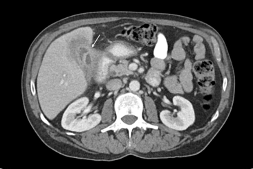
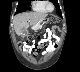
Over the next couple of days, John underwent extensive evaluation by the surgical oncology and gastroenterology team. A follow up CT, performed approximately 30 days after the initial exam, showed improvement in the gallbladder inflammation with resolution of air in the bile ducts, but persistent gallbladder wall thickening.

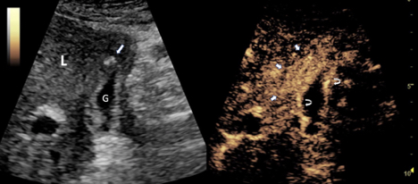
To address concerns of gallbladder cancer, the radiology team was asked to perform an ultrasound-guided biopsy of the gallbladder wall. Instead the radiologist recommended a contrast-enhanced ultrasound (CEUS) scan, since CEUS is a radiation-free imaging technique and gives instant results to aid in the decision for biopsy or other management of the gallbladder wall thickening. On a CEUS scan, gallbladder cancer would enhance differently when compared to inflammatory tissues.

Fortunately for John, CEUS images were not consistent with cancer; rather, the CEUS scan indicated only chronic gallbladder inflammation (Figure 3,4). As a result, John was a candidate for robotic cholecystectomy —  a much smaller procedure than extensive gallbladder surgery for cancer. Confirming the CEUS results, pathologic evaluation of the excised gallbladder wall showed no evidence of malignancy, but rather acute and chronic gallbladder inflammation.

Figure 3. Axial contrast-enhanced ultrasound (CEUS) of the gallbladder at a comparable level as CT images in figure 1, taken 2 weeks after the CT, on the left without contrast and on the right with contrast: The left image is optimized to show anatomy of liver (L) and gallbladder (G), the right image is optimized to show the contrast bubbles. One gallstone had migrated from the gallbladder into the liver (arrow on left). The irregular gallbladder wall thickening demonstrates strong enhancement (curved arrows on right) and does not demonstrate any underlying mass. There is strong enhancement of microbubbles in the liver parenchyma around the gallbladder (short arrows on right), corresponding to the dark zone of tissue on the CT (figure 1 and 2), which is typical for an inflammatory reaction due to gallbladder inflammation.

John’s experience shows how CEUS can help patients avoid unnecessary procedures and receive more appropriate care.  Thanks to the relatively simple CEUS scan, John was spared an unnecessary liver and gallbladder biopsy, and CEUS was critical for planning the uncomplicated laparoscopic gallbladder removal. John’s  postoperative course was uneventful and he has now fully recovered from the procedure — without any remaining symptoms.

Be Part of our Community

ICUS Membership is free.

ICUS Connect

FREE
VIEW