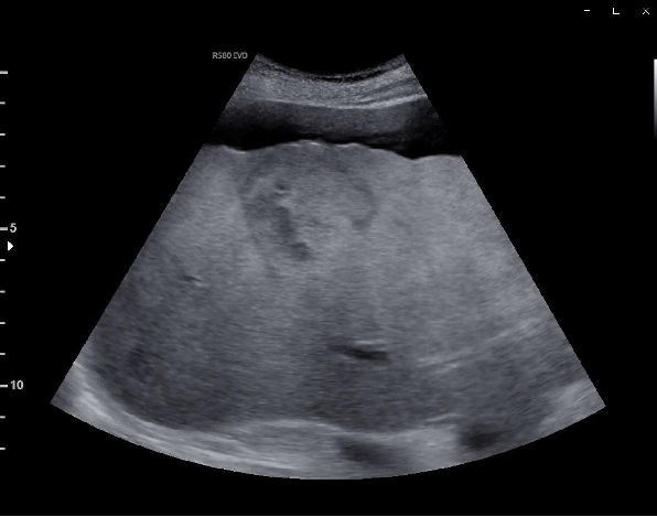
In the pre-existing liver cirrhosis, the baseline B-mode examination demarcates a heterogenic liver lesion, measuring 5.5 x 5.1 cm
The lesion shows no internal signal flow in the color Doppler and power Doppler examination.
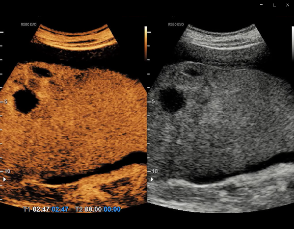
Following the intravenous administration of contrast agent, the lesion demonstrated peripheral nodular enhancement, progressing in a centripetal direction, areas of thrombosis and no washout during the entire examination
63-y/o male patient with clinical background of cirrhosis and ascites is referred to our department for a sonographic liver staging.
The typical CEUS feature of a hemangioma is peripheral, discontinuous nodular (syn.: globular) enhancement in the arterial phase with progressive centripetal partial or complete fill-in.
Complete fill-in occurs only in 40–50 % cases during the late phase. This filling-in is often faster in smaller lesions and the entire lesion may be hyperenhancing in the arterial phase
Choose your diagnosis:
Choose the final diagnosis:
CEUS is indicated when a definitive diagnosis of a hemangioma cannot be achieved using conventional US, as the addition of CEUS markedly improves the diagnostic accuracy in 90–95 % of the cases.
Overall sensitivity of CEUS for diagnosis of haemangioma is 86 % (95 %CI: 81–92 %).
CEUS is recommended for the characterization of focal liver lesions in the non-cirrhotic liver in patients with inconclusive findings at CT or MR imaging
The information, references and opinions provided on this website do not constitute the offering of medical advice or create a physician-patient relationship between any one or more of (a) the International Contrast Ultrasound Society (ICUS), its directors and members, and the contributors to this website, and (b) you, your patients, and any other party. ICUS makes no express or implied warranties regarding the accuracy or completeness of the information, references and opinions on this website, and ICUS shall not be liable to you, your patients, or anyone else with respect to any medical decision made, or any action taken or not taken, in direct or indirect reliance on this information.


International Contrast Ultrasound Society © 2025. All Rights Reserved.