NAFLD With no cirrhosis
48 y/o female
Surveillance revealed a liver mass that had increased in size from 1.4cm in 2018, to 4.5 cm in 2020.
She had no History of Birth Control use, and was sent to CEUS for further characterization of her liver mass.
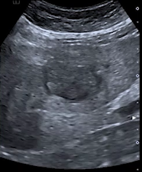
Greyscale image shows a large heterogeneous mass with a hypoechoic rim abutting the left portal vein
Color Doppler image shows distinct and prominent blood vessels centrally within the mass.
Choose your diagnosis:
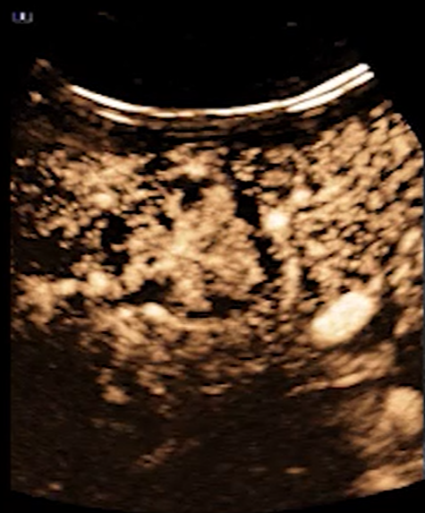
CEUS in the Arterial Phase (AP), from 0 to 11 seconds, shows avid enhancement within the mass with distinct centrifugal fill from the inside out.
CEUS in the Portal Venous Phase (PVP) at 5 mins suggests very weak washout.
Choose the final diagnosis:
Focal Nodular Hyperplasia (FNH) Benign Lesions are characterized by their distinct fill patterns in the Arterial Phase. Most benign lesions are found on surveillance ultrasound. CEUS can be done immediately, and it has exquisite spatial resolution making it outstanding for the visualization of the smallest of vessels especially with the early fill patterns of benign lesions. Although there may have been a suggestion of weak washout the fill pattern is unequivocal. The patient went on to more conservative management
The information, references and opinions provided on this website do not constitute the offering of medical advice or create a physician-patient relationship between any one or more of (a) the International Contrast Ultrasound Society (ICUS), its directors and members, and the contributors to this website, and (b) you, your patients, and any other party. ICUS makes no express or implied warranties regarding the accuracy or completeness of the information, references and opinions on this website, and ICUS shall not be liable to you, your patients, or anyone else with respect to any medical decision made, or any action taken or not taken, in direct or indirect reliance on this information.


International Contrast Ultrasound Society © 2025. All Rights Reserved.