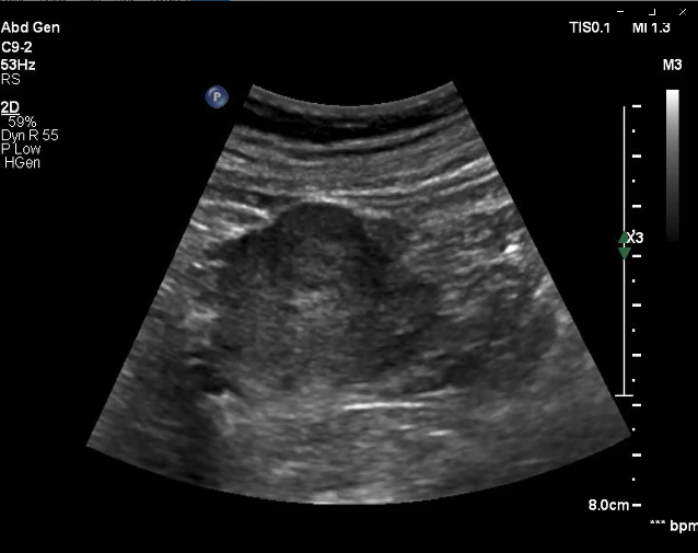
Gray-Scale B-Mode:
In the right kidney, a renal mass is demarcated with diameter of approximately 1,8 cm and slightly hyperechoic in comparison to the surrounding renal tissue. Color-coded duplex sonography depicted some vessels.
63 y/o female patient, came to the department due to suspicion of haemorrhage renal cyst in CT.
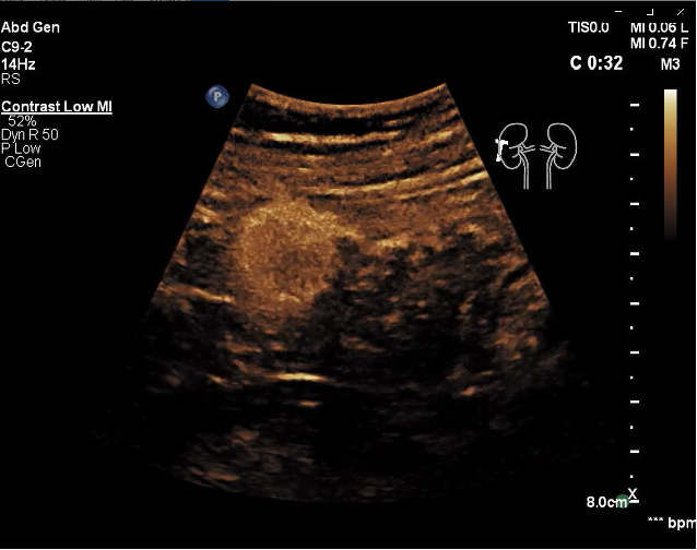
After intravenous ultrasound – contrast administration the lesion shows rapid contrast uptake in comparison to the renal parenchyma. Slight wash-out in the late phase was observed.
Choose your diagnosis:
Choose the final diagnosis:
Due to the contrast uptake a haemorrhage renal cyst could be excluded, afterwards the patient underwent biopsy and an oncozytoma was confirmed.
The information, references and opinions provided on this website do not constitute the offering of medical advice or create a physician-patient relationship between any one or more of (a) the International Contrast Ultrasound Society (ICUS), its directors and members, and the contributors to this website, and (b) you, your patients, and any other party. ICUS makes no express or implied warranties regarding the accuracy or completeness of the information, references and opinions on this website, and ICUS shall not be liable to you, your patients, or anyone else with respect to any medical decision made, or any action taken or not taken, in direct or indirect reliance on this information.
International Contrast Ultrasound Society © 2022. All Rights Reserved.
Created by customedialabs. Powered by momencio