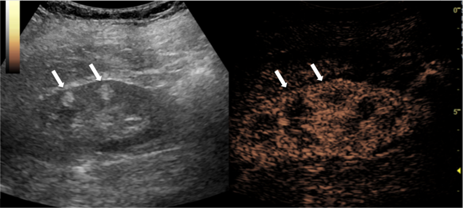
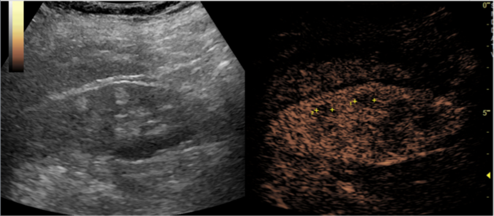
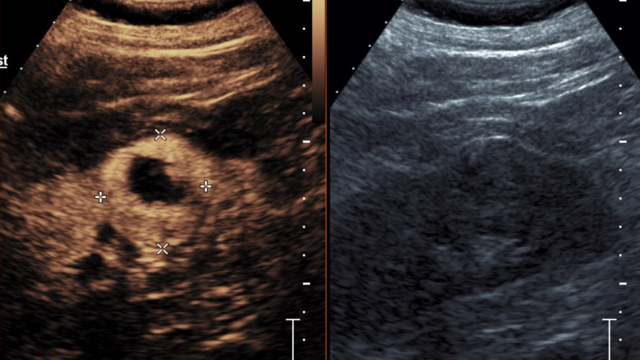
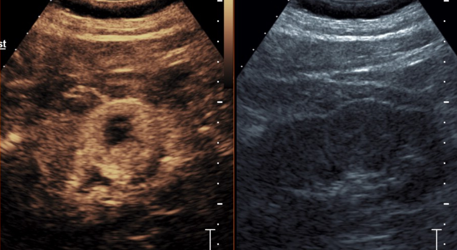
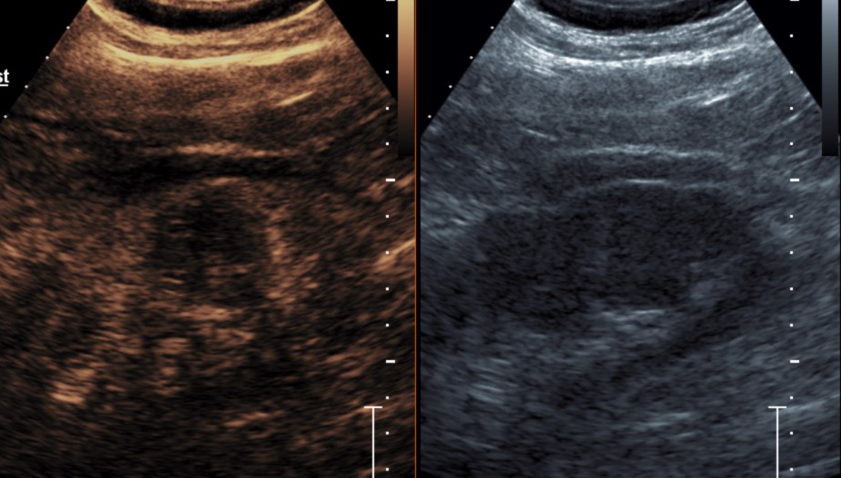
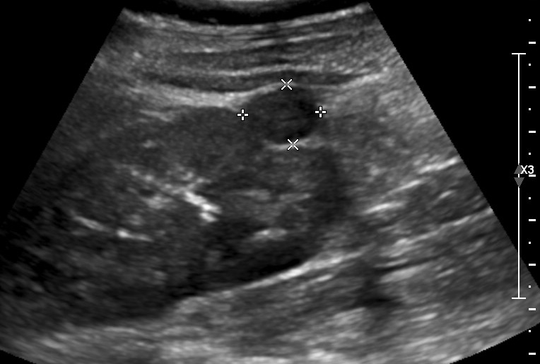
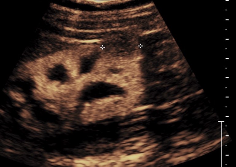
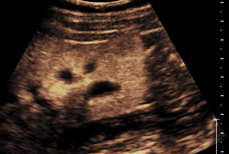
Imaging and timer begin with the start of the saline flush. Continuous recording from first arrival of a bubble in the FOV to lesion characterization, which usually less than 2- minutes. From the cine clips, still images can then be saved at the end of the study. Scanning the entire lesion particularly in indeterminate masses is required. Often scanning the kidney for additional lesions is possible.
CONTRAST AGENT DOSING: Depends on transducer and instrumentation, and body habitus. In general, for adult patients, a dose of 1.5-2.4 ml Lumason; Definity 0.2-0.4ml; Optison 1.0-1.5ml


- Barr, Richard G., et al., Society of Radiologists in Ultrasound (SRU): Contrast-enhanced Ultrasound—State of the Art in North America Ultrasound Quarterly: September 2020 – Volume 36 – Issue 4S – p S1-S39 doi: 10.1097/RUQ.0000000000000515
- Piscaglia, F., et al., The EFSUMB Guidelines and Recommendations on the Clinical Practice of Contrast Enhanced Ultrasound (CEUS): update 2011 on non-hepatic applications. Ultraschall Med, 2012. 33(1): p. 33-59.
- Sidhu PS., et.al. The EFSUMB Guidelines and Recommendations for the Clinical Practice of Contrast-Enhanced Ultrasound (CEUS) in Non-Hepatic Applications: Update 2017. Ultraschall in Med 2018; 39: e2–e44.
Barr RG. How to Develop a Contrast-Enhanced Ultrasound Program. J Ultrasound Med 2017.
Barr RG, Peterson C, Hindi A. Evaluation of indeterminate renal masses with contrast-enhanced US: a diagnostic performance study. Radiology 2014;271; 133-142.
Claudon M, Dietrich CF, Choi BI, Cosgrove DO, Kudo M, Nolsoe CP, Piscaglia F, Wilson SR, Barr RG, Chammas MC, Chaubal NG, Chen MH, Clevert DA, Correas JM, Ding H, Forsberg F, Fowlkes JB, Gibson RN, Goldberg BB, Lassau N, Leen EL, Mattrey RF, Moriyasu F, Solbiati L, Weskott HP, Xu HX. Guidelines and Good Clinical Practice Recommendations for Contrast Enhanced Ultrasound (CEUS) in the Liver – Update 2012. Ultraschall Med 2012.
Dietrich CF, Ignee A, Hocke M, Schreiber-Dietrich D, Greis C. Pitfalls and artefacts using contrast enhanced ultrasound. Z Gastroenterol 2011;49; 350-356.
Fetzer DT, Rafailidis V, Peterson C, Grant EG, Sidhu P, Barr RG. Artifacts in contrast-enhanced ultrasound: a pictorial essay. Abdom Radiol (NY) 2018;43; 977-997.
O’Neal D, Cohen T, Peterson C, Barr RG. Contrast-Enhanced Ultrasound-Guided Radiofrequency Ablation of Renal Tumors. J Kidney Cancer VHL 2018;5; 7-14.
Piscaglia F, Nolsoe C, Dietrich CF, Cosgrove DO, Gilja OH, Bachmann Nielsen M, Albrecht T, Barozzi L, Bertolotto M, Catalano O, Claudon M, Clevert DA, Correas JM, D’Onofrio M, Drudi FM, Eyding J, Giovannini M, Hocke M, Ignee A, Jung EM, Klauser AS, Lassau N, Leen E, Mathis G, Saftoiu A, Seidel G, Sidhu PS, ter Haar G, Timmerman D, Weskott HP. The EFSUMB Guidelines and Recommendations on the Clinical Practice of Contrast Enhanced Ultrasound (CEUS): update 2011 on non-hepatic applications. Ultraschall Med 2012;33; 33-59.
Tenant SC, Gutteridge CM. The clinical use of contrast-enhanced ultrasound in the kidney. Ultrasound 2016;24; 94-103.
Sparchez, Z., et al., Contrast enhanced ultrasound of renal masses. A reappraisal of EFSUMB recommendations and possible emerging applications. Med Ultrason, 2015. 17(2): p. 219-26.
Sidhu, P.S., et al., The EFSUMB Guidelines and Recommendations for the Clinical Practice of Contrast-Enhanced Ultrasound (CEUS) in Non-Hepatic Applications: Update 2017 (Long Version). Ultraschall Med, 2018. 39(2): p. e2-e44